Pathophysiology Specific structural, functional, and neurotransmitter changes in the brain may be associated with ADHD Specific structural, functional, and neurotransmitter changes in the brain may be associated with ADHD
Structural
Structural imaging studies include volumetric measurements of gray or white matter of the whole brain (including or excluding the cerebellum) and its lobes, and fine-grained measurements (e.g., cortical thickness, density of gray matter) acquired from individual voxels in the brain or across the cortical surface.19Adapted from Jadidian A, Hurley RA, Taber KH. Neurobiology of adult ADHD: Emerging evidence for network dysfunctions. J Neuropsychiatry Clin Neurosci. 2015;27(3):173-178.
Adapted from Jadidian A, Hurley RA, Taber KH. Neurobiology of adult ADHD: Emerging evidence for network dysfunctions. J Neuropsychiatry Clin Neurosci. 2015;27(3):173-178.
Individuals with ADHD have structural differences in their brains compared with individuals without ADHD
Gray matter density is lower20,21
Localized volumetric gray matter abnormalities in the basal ganglia20
Reduced right globus pallidus and putamen volumes and decreased caudate volumes in manual tracing studies in children with ADHD20
Volume reduction in the anterior cingulate cortex of adults with ADHD20
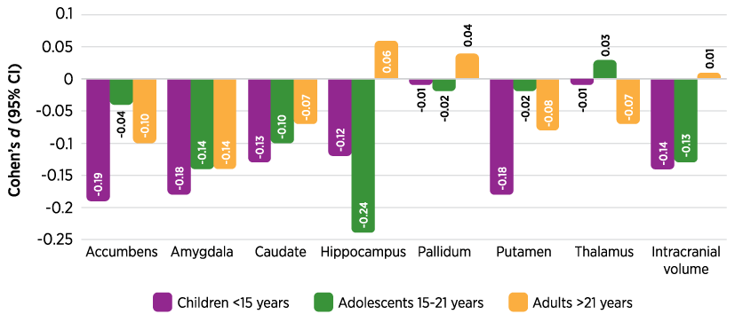
Smaller volume overall and in specific structures19,20,23
Large MRI study showed volumes of various brain structure were slightly smaller in children, adolescents, and adults with ADHD (N=1,713) compared with controls (N=1,529)23
White matter abnormalities
Lobar white matter volume reduced ~4% in children and adolescents (N=152) with ADHD aged 5-18 years19,22
Abnormally high fractional anisotropy in frontal networks in adolescents (N=14) with ADHD19
Cortical differences
Delayed cortical maturation in children and adolescents (N=223) aged 7-13 years24
Reduced cortical thickness in adults.20,25

Functional changes
Regions of the brain that are associated with ADHD correspond to networks involving frontal regions, executive function, and attention.26
Functional neuroimaging studies have shown variation in activation/suppression of networks in ADHD
– Under-activation of frontostriatal and frontoparietal circuits, and other frontal brain regions20,27-29
– Under-activation of systems involved in executive function and attention34,44
– Over-activation (reduced suppression) of the default mode network during task performance30,31
Neurotransmitter changes
Neuromodulatory influences over catecholamines in the fronto-striato-cerebellar regions play important roles in high-level executive functions32
ADHD symptoms may be related to dysregulation of basal, tonic catecholaminergic levels30,33
– Too low: distractibility
– Too high: hyperactivity and anxiety
Dopamine18,30,34

– Deficit in dopaminergic signaling30
Noradrenaline

– Noradrenergic signaling34,35
Glutamate36,37

– Levels of glutamate/glutamine are significantly
reduced in the caudate/putamen of adults with
ADHD compared to adults without ADHD.
